
Full Body MRI: The Gold Standard for Preventive Screening
Why radiation-free whole-body MRI has become a foundational tool in preventive medicine — what it screens for, how it works, and who it's for.
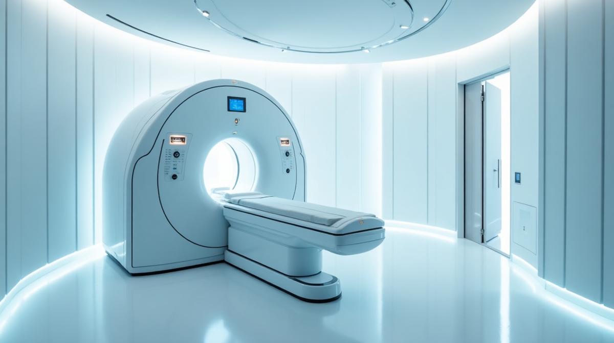
Full-body magnetic resonance imaging (MRI) has moved from a specialty tool used for staging known disease to a foundational instrument in modern preventive and longevity medicine. Unlike CT scans and X-rays, full-body MRI uses no ionizing radiation. Instead, it relies on powerful magnetic fields and radio-frequency pulses to produce extraordinarily detailed images of soft tissue, organs, vasculature, and the musculoskeletal system.
For individuals focused on extending healthspan and detecting disease at its earliest, most treatable stage, a high-quality full-body MRI offers something most other screening tools cannot: a single, comprehensive look at the body without exposing the patient to cumulative radiation risk. At ReGenesis Longevity Clinic™, full-body MRI is one of the cornerstone screening tools recommended within a comprehensive longevity assessment.
What Full-Body MRI Actually Looks At
A modern full-body MRI protocol typically images the brain, neck, spine, chest, abdomen, pelvis, and major joints. Depending on the protocol and indication, the study can also include dedicated sequences for the prostate, pelvic organs, vasculature, and musculoskeletal areas of concern. The goal is breadth — covering organ systems and structures that are not routinely screened by conventional medicine.
Across these regions, MRI is well suited to detecting solid organ masses, cystic lesions, aneurysms, areas of unexpected inflammation, structural abnormalities of the brain and spine, soft-tissue tumors, certain types of cancer, and joint or disc pathology. It is not, however, a complete answer to every screening question; it is one component within a multi-modality preventive strategy.
Why MRI — and Not CT or X-ray?
The single most important advantage of MRI is that it uses no ionizing radiation. This matters significantly for screening, where the same patient may undergo repeated studies over decades. Cumulative radiation exposure from CT scans is well-documented to carry a small but real long-term cancer risk, particularly in younger patients and those undergoing repeated imaging. MRI sidesteps that concern entirely.
MRI also offers superior soft-tissue contrast. This makes it the imaging modality of choice for the brain, spinal cord, liver, pancreas, kidneys, prostate, pelvic organs, and musculoskeletal soft tissue. Many early lesions that are difficult to characterize on CT are clearly visible on MRI, which is one of the main reasons it has become the preferred radiation-free screening tool.
What a Full-Body MRI Can Help Detect Early
Early detection is where preventive imaging earns its place. Solid organ tumors of the liver, kidneys, pancreas, and ovaries — many of which are typically diagnosed at later, harder-to-treat stages — can sometimes be identified incidentally on a high-quality full-body MRI. Brain aneurysms, white matter changes, structural brain lesions, and spinal abnormalities are all visible on the same study.
MRI is also exceptionally good at characterizing musculoskeletal pathology: rotator cuff tears, hip joint pathology, lumbar disc disease, sacroiliac inflammation, and meniscal injuries can be identified, often clarifying chronic pain that has not been adequately explained by clinical examination alone.
Limitations and What MRI Does Not Replace
It is important to be precise about what full-body MRI is — and is not. It is an exceptionally good screening tool for many soft-tissue and structural pathologies, but it is not a substitute for every other test. Mammography remains the standard for breast cancer screening; colonoscopy remains the standard for colorectal screening; coronary artery calcium scoring and dedicated cardiac imaging address cardiovascular risk in ways MRI cannot. Lung nodule surveillance is generally better suited to low-dose CT in appropriate populations.
A well-designed preventive program uses full-body MRI as part of a layered strategy: comprehensive blood work, cardiovascular risk stratification, disease-specific screening, and targeted imaging where indicated. The MRI is one essential layer — not the entire structure.
Incidental Findings: A Real Consideration
Whole-body imaging frequently detects incidental findings — small cysts, benign nodules, or non-specific changes that may have no clinical significance. Without thoughtful interpretation, these can lead to unnecessary follow-up testing, anxiety, and procedures. This is one of the reasons it is critical that full-body MRI be ordered, interpreted, and contextualized within a clinically supervised longevity program rather than treated as a standalone consumer product.
At ReGenesis, every imaging study is reviewed in the context of the patient's complete clinical picture. Findings that warrant follow-up are addressed with appropriate next steps; findings that are clearly benign are documented but do not trigger over-investigation.
Who Benefits Most from Full-Body MRI
Full-body MRI is particularly well-suited to adults focused on long-term health optimization, individuals with a meaningful family history of cancer or cardiovascular disease, executives and professionals with limited time for fragmented testing, and patients who want a structured baseline of their internal anatomy that can be re-imaged in future years to detect change.
It is generally less indicated in patients with uncontrolled symptoms requiring acute work-up; in those cases, targeted imaging directed by clinical findings is the appropriate path. As with any screening modality, the decision to scan should be individualized and integrated into a broader assessment.
How ReGenesis Uses Full-Body MRI
Within the ReGenesis longevity assessment, full-body MRI is offered as part of a comprehensive baseline. The imaging is interpreted in conjunction with comprehensive blood work, cardiovascular risk markers, body composition, and lifestyle assessment. The result is not a stack of unread images, but a clinically synthesized picture of where the patient stands and what the most important next steps are.
Used this way, full-body MRI becomes one of the most powerful tools available in preventive medicine: a high-resolution, radiation-free look at the body, integrated into a longitudinal plan designed to detect disease early, monitor change over time, and support a longer, healthier life.
How a Full-Body MRI Appointment Actually Works
From the patient's perspective, a full-body MRI is a relatively simple experience. After a brief intake and safety screening — particularly important for ruling out incompatible implants, devices, or metal — the patient changes into a gown and lies on the scanner table. The scan is non-invasive and painless, although the magnet is loud and the experience requires lying still for an extended period. Most full-body protocols take roughly 60 to 90 minutes.
Patients who experience claustrophobia can usually be accommodated through wider-bore scanners, careful preparation, and in some cases, mild anxiolytic medication arranged in advance with their clinician. Communication with the technologist is continuous throughout the study, and the patient can stop the scan at any time.
Establishing a Baseline for Future Comparison
One of the most underappreciated benefits of a high-quality full-body MRI is the value of having a baseline. Many findings on imaging are difficult to interpret in isolation: a small lesion, a mild signal change, or a borderline structural finding may be entirely benign and stable, or may be the earliest sign of evolving pathology. Without a prior study for comparison, it is often impossible to tell.
Establishing a clear baseline in midlife and following it forward over time gives clinicians the ability to detect change rather than simply reacting to a snapshot. A finding that has been stable for years on serial imaging is reassuring; a finding that is new or evolving warrants attention. Serial comparison transforms imaging from a one-time event into a longitudinal tool.
Patient Experience and Reporting
Beyond the scan itself, the value of a full-body MRI program depends heavily on the quality of the radiology read and the way findings are communicated. A high-quality program ensures that subspecialty radiologists review the images, that findings are reported with clear language and clinical context, and that the patient receives a structured review of the results with their longevity provider — not simply a copy of a technical report.
At ReGenesis, every imaging study is integrated into the patient's broader longevity assessment. Findings are explained in plain language, contextualized against the patient's overall risk profile, and translated into a clear plan: monitor, investigate further, or take no further action. That clinical translation is what turns imaging data into useful health information.
Frequently Asked Questions About Full-Body MRI
How often should a full-body MRI be repeated?
For most adults pursuing full-body MRI as part of a longevity program, a baseline study followed by repeat imaging at 1- to 3-year intervals is reasonable. Higher-risk individuals — significant family history, known surveillance lesions, or specific clinical concerns — may warrant more frequent imaging. The cadence is individualized based on the patient's overall risk profile, prior findings, and goals.
Is there any radiation exposure?
No. MRI uses magnetic fields and radio-frequency pulses to generate images and does not involve ionizing radiation. This is one of the central reasons it is favoured for repeat preventive imaging, particularly in patients who may undergo serial studies over many years.
What about contrast dye?
Many full-body screening protocols are performed without intravenous contrast. When contrast is required for a specific clinical question, gadolinium-based contrast agents are used and are very safe in patients with normal kidney function. The decision to use contrast is made based on the indication and the patient's individual situation.
Will it find everything?
No imaging study finds every possible condition. Full-body MRI is exceptionally good at detecting many soft-tissue and structural pathologies, but it does not replace dedicated screening for breast cancer, colon cancer, or coronary artery disease. It works best as one important component of a layered, individualized preventive strategy.
Where Full-Body MRI Fits in a Modern Preventive Strategy
Full-body MRI is most powerful when it is integrated into a broader preventive program rather than treated as a standalone scan. The most thoughtful approach pairs the MRI with comprehensive blood work, advanced cardiovascular risk assessment (including markers such as ApoB and lipoprotein(a) where appropriate), structured cancer screening per established guidelines, and longitudinal clinical follow-up. Each of these elements addresses a different dimension of risk, and the combination is what produces a genuinely complete preventive picture.
Within that framework, full-body MRI plays a specific and valuable role: comprehensive structural assessment, radiation-free, with the soft-tissue resolution required to detect many early lesions, vascular abnormalities, and structural changes that other modalities miss. It is not the answer to every preventive question, but it is one of the most important answers to many of them — and its value compounds over time as serial studies allow change to be detected with high confidence.
For patients who are serious about long-term health, full-body MRI is increasingly considered an essential part of a high-quality longevity assessment. At ReGenesis, it is offered within exactly that broader program — comprehensive, individualized, and longitudinal — so that imaging data becomes one important input in a complete healthspan strategy rather than an isolated event.
For patients serious about long-term health, full-body MRI offers something few other single tools can: a comprehensive, radiation-free structural baseline that can be revisited and compared over years. Combined with comprehensive blood work and structured longevity follow-up, it becomes one of the most valuable components of a modern preventive program.